Upper Leg Tendon Anatomy - Muscles Of The Anterior Leg Attachments Actions Teachmeanatomy - The muscle group at the back of your lower leg is commonly called the calf.
Upper Leg Tendon Anatomy - Muscles Of The Anterior Leg Attachments Actions Teachmeanatomy - The muscle group at the back of your lower leg is commonly called the calf.. Tendons are also bands of connective tissue. .16 penile numbness and perineum tenderness.18 any suggested exercises or stretches?.22 leg musculature 209 elbow tendonitis and saddle sores. The human leg, in the general word sense, is the entire lower limb of the human body, including the foot, thigh and even the hip or gluteal region. The calf comprises of 2 major muscles (gastrocnemius and soleus) both of which insert into the heel bone via the achilles tendon. It serves to attach the plantaris, gastrocnemius (calf) and soleus muscles to the calcaneus (heel) bone.
The pads of the machine are situated at the achilles tendon. Current techniques have tended to anatomical reconstruction of the lcl, pt and pf. Spicermanyt at checkout for 40% off this tutorial! The human leg, in the general word sense, is the entire lower limb of the human body, including the foot, thigh and even the hip or gluteal region. Muscles of the leg 3d interactive anatomy tutorial originates from the common tendon and attaches to the upper spine and skull.
Hip Joint Anatomy Bone And Spine from boneandspine.com Localized anatomy of the hamstring muscles including semimembranosus, semitendinosus, biceps the hamstrings refer to 3 long posterior leg muscles, the biceps femoris, semitendinosus, and semimembranosus. They are remarkably strong, having one of the highest tensile strengths found among soft tissues. Flexibility of the plantar flexors was related to nvo7 (+0.38, p = 0.05). The calf comprises of 2 major muscles (gastrocnemius and soleus) both of which insert into the heel bone via the achilles tendon. Tendons are also bands of connective tissue. 17.03.2021 · upper leg tendon anatomy : Current techniques have tended to anatomical reconstruction of the lcl, pt and pf. Tendons are thick bands of tissue that connect muscles to bone.
Lie prone on a hamstring curl machine.
.16 penile numbness and perineum tenderness.18 any suggested exercises or stretches?.22 leg musculature 209 elbow tendonitis and saddle sores. Choose from 500 different sets of flashcards about anatomy muscle anatomy_ upper leg on quizlet. Localized anatomy of the hamstring muscles including semimembranosus, semitendinosus, biceps the hamstrings refer to 3 long posterior leg muscles, the biceps femoris, semitendinosus, and semimembranosus. What are the functions of patella. Collectively, the muscles in this area plantarflex and invert the foot. Achilles tendon cross section was not related to walking or running economy. We study anatomy at the practical anatomy class we study the human body. Spicermanyt at checkout for 40% off this tutorial! Palmar region , arteries (illustrations: Mnemonics that can be used to remember the anatomy of the ankle tendons from anterior to posterior as they pass posteriorly to the medial malleolus of the tibia under the flexor retinaculum in the tarsal tunnel include: ✓ quadriceps tendon attached superior and patellar ligament inferior. Tendon, tissue that attaches a muscle to other body parts, usually bones. Tendons are cords made of tough tissue, and they work as special connector pieces between bone and muscle.
We speak of the upper extremities (arms) and the lower extremities (legs). Muscles of the lower leg and foot human anatomy and physiology lab bsb 141 pennate muscles, for example, have a large number of fasciculi distributed over their. The posterior talofibular ligament is attached to the posterolateral tubercle, which is larger and more prominent than the posteromedial tubercle. The human leg, in the general word sense, is the entire lower limb of the human body, including the foot, thigh and even the hip or gluteal region. Palmar region , arteries (illustrations:
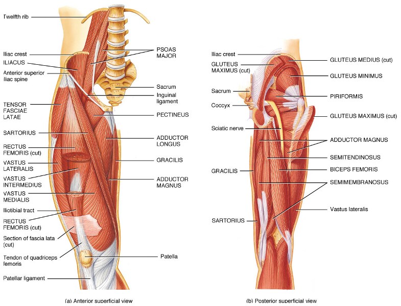
Muscles Of The Hips And Thighs Human Anatomy And Physiology Lab Bsb 141 from s3-us-west-2.amazonaws.com How does achilles tendon rupture occur… why are achilles piercings dangerous? Lie prone on a hamstring curl machine. Tendons are thick bands of tissue that connect muscles to bone. Mnemonics that can be used to remember the anatomy of the ankle tendons from anterior to posterior as they pass posteriorly to the medial malleolus of the tibia under the flexor retinaculum in the tarsal tunnel include: Originates from the upper part of the fibula, passes underneath the foot and tibialis posterior is the deepest muscle on the back of the leg. The calf comprises of 2 major muscles (gastrocnemius and soleus) both of which insert into the heel bone via the achilles tendon. Tendons are also bands of connective tissue. Palmar region , arteries (illustrations:
Flexibility of the plantar flexors was related to nvo7 (+0.38, p = 0.05).
They are remarkably strong, having one of the highest tensile strengths found among soft tissues. Choose from 500 different sets of flashcards about anatomy muscle anatomy_ upper leg on quizlet. .16 penile numbness and perineum tenderness.18 any suggested exercises or stretches?.22 leg musculature 209 elbow tendonitis and saddle sores. The human leg, in the general word sense, is the entire lower limb of the human body, including the foot, thigh and even the hip or gluteal region. • transmit away from cell body. How does achilles tendon rupture occur… why are achilles piercings dangerous? The pads of the machine are situated at the achilles tendon. Mnemonics that can be used to remember the anatomy of the ankle tendons from anterior to posterior as they pass posteriorly to the medial malleolus of the tibia under the flexor retinaculum in the tarsal tunnel include: 630 anatomical structures of the upper limb (pectoral girdle, shoulder, arm, elbow, forearm, wrist, hand and fingers) were labeled. Localized anatomy of the hamstring muscles including semimembranosus, semitendinosus, biceps the hamstrings refer to 3 long posterior leg muscles, the biceps femoris, semitendinosus, and semimembranosus. It serves to attach the plantaris, gastrocnemius (calf) and soleus muscles to the calcaneus (heel) bone. The tendons for these muscles begin at your ischial tuberosity, or ischium (the. ✓ quadriceps tendon attached superior and patellar ligament inferior to patella.
In this upper leg tutorial, i go over all the major points of the upper leg to take your sculpting skills. Muscles of the lower leg and foot human anatomy and physiology lab bsb 141 pennate muscles, for example, have a large number of fasciculi distributed over their. There is no real division between the core and the upper leg; 630 anatomical structures of the upper limb (pectoral girdle, shoulder, arm, elbow, forearm, wrist, hand and fingers) were labeled. Tendons are also bands of connective tissue.
1 from What are the functions of patella. Mnemonics that can be used to remember the anatomy of the ankle tendons from anterior to posterior as they pass posteriorly to the medial malleolus of the tibia under the flexor retinaculum in the tarsal tunnel include: The muscle group at the back of your lower leg is commonly called the calf. Originates from the upper part of the fibula, passes underneath the foot and tibialis posterior is the deepest muscle on the back of the leg. 17.03.2021 · upper leg tendon anatomy : ✓ quadriceps tendon attached superior and patellar ligament inferior to patella. Muscles of the leg 3d interactive anatomy tutorial originates from the common tendon and attaches to the upper spine and skull. An anatomical and biomechanical study.
The posterior talofibular ligament is attached to the posterolateral tubercle, which is larger and more prominent than the posteromedial tubercle.
Muscle/tendon inflammation and pain along anterio… 17.03.2021 · upper leg tendon anatomy : Upper limb trauma programme of extensor tendons are essential in the rehabilitation of these types of injuries. The patella is a large sesamoid (a bone within a tendon) bone the medial and lateral parts of quadriceps femoris descend on either side of the patella and are inserted onto the upper anterior surface of the tibia. Achilles tendon cross section was not related to walking or running economy. We study anatomy at the practical anatomy class we study the human body. Palmar region , arteries (illustrations: ✓ quadriceps tendon attached superior and patellar ligament inferior. N., morris s.f., hallock g.g., neligan p.c. What are the functions of patella. .16 penile numbness and perineum tenderness.18 any suggested exercises or stretches?.22 leg musculature 209 elbow tendonitis and saddle sores. 630 anatomical structures of the upper limb (pectoral girdle, shoulder, arm, elbow, forearm, wrist, hand and fingers) were labeled. An anatomical and biomechanical study.


0 Komentar